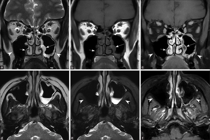
Figure 2.
Normal maxillary sinus on the right side and chronic noninvasive maxillary sinusitis on the left side Coronal T2W (a), T1W (b), postcontrast T1W image with fat saturation (c), axial T2W (d), T2W with fat saturation (e), and postcontrast T1W image with fat saturation (f). Note the normal mucosa in the right maxillary sinus (short arrow) and thickened, peripherally enhancing mucosa in the left maxillary sinus (long arrow). Note the absence of edema/enhancement in the bony structures and periantral fat (arrow heads)

