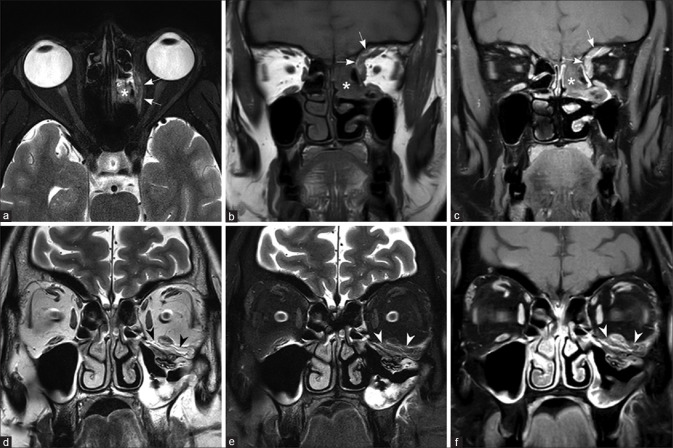
Figure 5.
Orbital extension from ethmoid sinus (a-c) and maxillary sinus (d-f) Fat-saturated T2W (a), T1W (b), and fat-saturated post-contrast T1W images (c) in a patient with early orbital extension shows the left ethmoid sinusitis (asterisk), with a thin enhancing extraconal soft tissue, involving the medial rectus, superior rectus, and superior oblique muscles (arrows). T2W (d), fat-saturated T2W (e), and fat-saturated postcontrast T1W images (f) in another patient show the left maxillary sinusitis with extension along the floor of the left orbit, involving the inferior rectus muscle (arrowheads)

