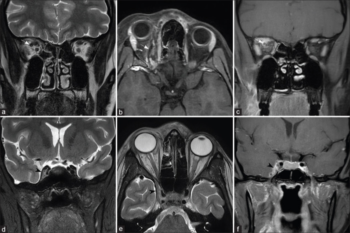
Figure 9.
Involvement of the superior ophthalmic vein and cavernous sinus. The right superior ophthalmic vein shows increased caliber with retroorbital fat stranding on T2W image (a) and filling defects on fat-saturated postcontrast T1W (b and c) images (arrows). T2W (d and e) sections through the cavernous sinuses show bulky right cavernous sinus with convex lateral wall (arrowheads) and filling defects on postcontrast T1W image (f)

