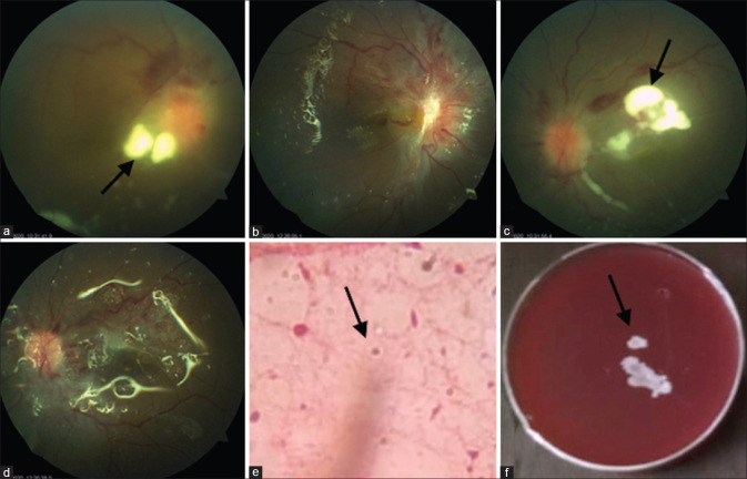
Figure 1.
Bilateral involvement in case 1. (a and c) Right and left eyes, respectively, showing whitish fluffy lesions (arrows) at the posterior pole, breakthrough preretinal hemorrhage, and mild disc hyperemia. (b and d) Postvitrectomy picture in the right and left eyes, with silicone oil in situ, clear vitreous cavity, and small fibrous proliferation over the optic disc. (c) Wet film 10% KOH mount shows double-walled yeasts-like organism. (arrow) (f) Growth on blood agar is seen as creamy white confluent colonies of Candida sp

