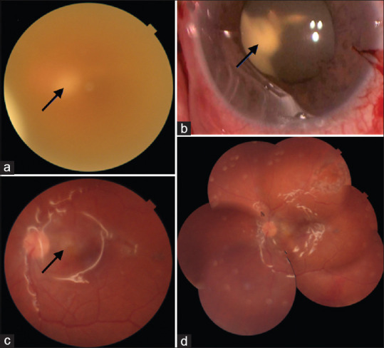
Figure 2.

Left affected eye in case 3. (a) Fundus photograph showing whitish retinal abscess (arrow) seen hazily through the turbid vitreous. (b) Intraoperatively, a large clump of exudates (arrow) was seen in the retrolenticular space. Lensectomy and retinotomy were necessary to remove this large granuloma. (c) 1 posttreatment, the retinal view improved and the resolving retinal abscess is seen (arrow). (d) Fundus photo montage shows clear vitreous cavity, silicone oil in situ, and well-attached retina
