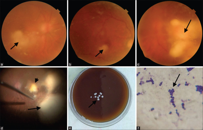
Figure 3.
Bilateral involvement in case 5. (a) Right eye showed diffuse fluffy cotton balls and a retinal abscess (arrow). (b) four weeks postvitrectomy; the vitreous cavity is clear, and the retinal abscess (arrow) is resolving. (c) the worse affected left eye showed a large clump of vitreous exudates (arrow). (d) intraoperatively a retinal abscess is seen at the posterior pole (arrowhead) along with two larger retinal abscesses (arrow). (e) Blood agar showing creamy white, smooth, discrete, and well-defined colonies of Candida sp. (arrow) (f) Gram stain showing budding gram-positive yeast cells. (arrow)

