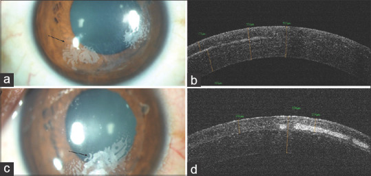
Figure 9.

Slit-lamp photograph of (a) the right eye two years after LASIK showing white opacity extending up to 2 mm inside from the flap edge suggestive of grade 2 epithelial ingrowth; (b) ASOCT image of the same eye showing increased flap thickness with interface hyperreflectivity; (c) the left eye with white opacity extending beyond 2 mm of the flap edge suggestive of grade 3 epithelial ingrowth; (d) ASOCT image of the same eye showing increased flap thickness with interface hyperreflectivity
