
Figure 1.
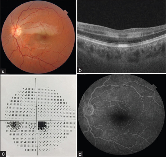
(a) Color fundus photograph of the isolated cilioretinal artery occlusion at the time of admission. (b) Paracentral acute middle maculopathy image on optical coherence tomography. (c) Paracentral scotoma on standard automated perimetry. (d) Filling delay in the cilioretinal artery in fluorescein angiography. The filling of the cilioretinal artery started at 21 s