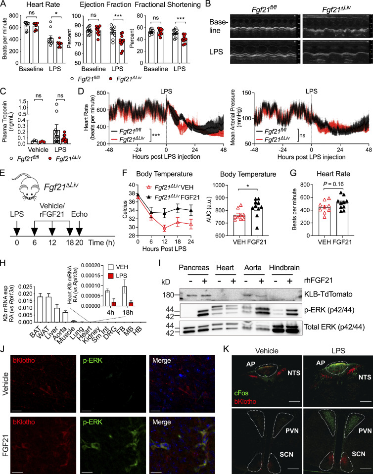
Figure 4.
FGF21 deficiency causes bradycardia during LPS endotoxemia. (A and B) Echocardiography performed on Fgf21fl/fl and Fgf21ΔLiv mice before (baseline) and 18 h after 5 mg/kg i.p. LPS. n = 10/group; pooled data are from three independent experiments. (B) Representative echocardiogram windows. (C) Plasma troponin in Fgf21fl/fl and Fgf21ΔLiv mice 20 h after PBS vehicle or 5 mg/kg i.p. LPS. Vehicle n = 3/group; LPS n = 10–12/group; pooled data are from three independent experiments. (D) Ambulatory blood pressure and heart rate of Fgf21fl/fl and Fgf21ΔLiv mice measured by in vivo telemetry before and after 2 mg/kg i.p. LPS. n = 4/group; pooled data are from three independent experiments. Mean arterial pressure and heart rate measured every minute and shown as mean ± SEM within each group. (E–G) Fgf21ΔLiv mice were challenged with LPS 5 mg/kg i.p. Recombinant mouse FGF21 10 ng i.v. or PBS vehicle was administered starting at 6 h after LPS and then every 6 h for three doses. n = 9–10/group; pooled data are from two independent experiments. Experimental workflow shown in E. (F) Rectal temperatures and areas under the curve (AUC). (G) Heart rate by echocardiography was performed at 20 h after LPS injection. (H) Klb mRNA expression in whole tissue from WT mice, shown relative to Rpl13a; inset shows RA of heart Klb after PBS vehicle or LPS treatment. n = 3–4/group; results of one of two independent experiments with similar results are shown. qPCR Ct values are as follows: brown adipose tissue (BAT), 22; white adipose tissue (WAT), 22; liver, 23; aorta, 25; heart, 30 at baseline, 32 after LPS. FB, forebrain; MB, midbrain; HB, hindbrain; Sm Int, small intestine; DRG, dorsal root ganglia. (I) Whole-tissue protein lysates from KlbTdTm mice immunoblotted for RFP (to detect KLB-TdTomato), p-ERK, and total ERK 10 min after 1 mg/kg i.p. recombinant human FGF21 (rhFGF21). The hindbrain region, including AP and NTS, was grossly dissected. Pancreas tissue was included as a positive control. Representative blots from one of three independent experiments are shown. (J) Brains from KlbTdTm mice were harvested 10 min after 1 mg/kg i.p. rhFGF21 treatment. 50-micron fixed brain vibratomed sections were immunostained for RFP and p-ERK. Representative images of the AP from one of two independent experiments are shown. Scale bars represent 20 µm. (K) Brains from KlbTdTm mice were harvested 18 h after 15 mg/kg i.p. LPS or PBS vehicle. 50-micron fixed brain vibratomed sections were immunostained for RFP and cFos. Representative images from one of three independent experiments are shown. PVN, paraventricular nucleus. Scale bars represent 200 µm. *, P < 0.05; ***, P < 0.001; two-way ANOVA with Sidak’s multiple comparisons test (A and C), two-way ANOVA (D), or unpaired two-sided t test (F and G). Data are expressed as mean ± SEM.

