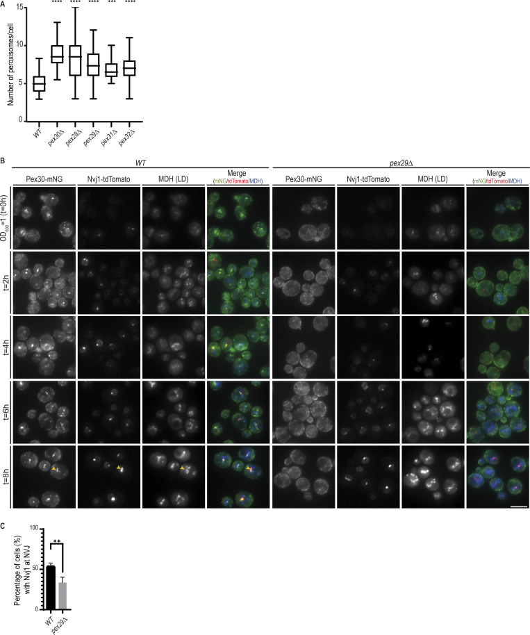
Figure S4.
Pex30/Pex29 role on NVJ formation. (A) Quantification of the number of peroxisomes per cell, in exponentially growing cells. Three independent experiments were analyzed (>30 cells/genotype/experiment were counted). Box and whiskers represent distribution of the values (minimum, 25th percentile, median, 75th percentile, and maximum). Ordinary one-way ANOVA and Dunnett’s multiple comparisons were done to compare the number of peroxisomes with the WT condition (****, P < 0.0001; ***, P < 0.001). (B) Time-course analysis of Pex30-mNG distribution in WT and pex29Δ cells grown in synthetic medium. Exponentially growing cells were monitored through diauxic shift into early stationary phase as indicated. The formation of NVJ was monitored by endogenous Nvj1-tdTomato, and LDs were stained with the neutral lipid dye MDH. Yellow arrowheads indicate cells with NVJ-clustered LDs. (C) Quantification of cells with the indicated genotype displaying Nvj1 localized to the NVJ during diauxic shift (4 h). Three independent experiments were analyzed (>30 cells/genotype/experiment were counted). Bars represent SD. Ordinary one-way ANOVA and Dunnett’s multiple comparisons were done to compare the percentage of cells with the WT condition (**, P < 0.01).

