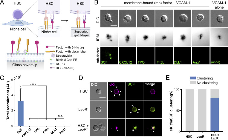
Figure 1.
mSCF is the only factor recruited/clustered by HSCs in a screening. (A) Illustration of membrane-bound HSC–niche cell interaction and recapitulation on an SLB. (B) HSCs after incubation with VCAM-1 and a membrane-bound factor (mb-factor; with fluorescent label, background subtracted) on SLBs for 1 h. DIC, differential interference contrast. Dark regions represent cell–substrate contact/adhesion. Scale bar = 5 µm. (C) Total membrane-bound factors recruited by single HSCs assessed by the total fluorescence under each cell. n = 37 cells per condition. Error bars represent SD. ****, P < 0.0001 by one-way ANOVA with Tukey’s test. AU, arbitrary units. (D) HSCs, LepR+ MSCs, and HSC–LepR+ MSC pairs forming physical contact after 1-h incubation. Arrowheads point to dispersed cKit or SCF on the surface of single HSCs or LepR+ MSCs, respectively, or clustered SCF/cKit at the HSC–MSC interface. Scale bar = 5 µm. (E) Frequency of cKit/mSCF clustering on one side of the cell in single cells or in HSC–MSC pairs. n = 20 single cells or cell pairs for each condition.

