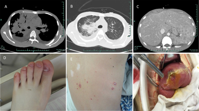
Figure 1.
Clinical photos. (A) Bilateral lung infection with a bilateral pleural effusion, a paraspinal mass of the right lower mediastinum, and pericardial effusion. (B) CT of pulmonary Talaromyces marneffei infection. (C) Enlarged spleen, multiple splenic lesions, cholecystitis, and peritoneal effusion. (D) Abnormality of the patient's toenails. (E) Rash on the patient's back due to Talaromyces marneffei infection. (F) Surgical exploration demonstrated intestinal perforation and edema of the surrounding mucosa.

