Figure 5.

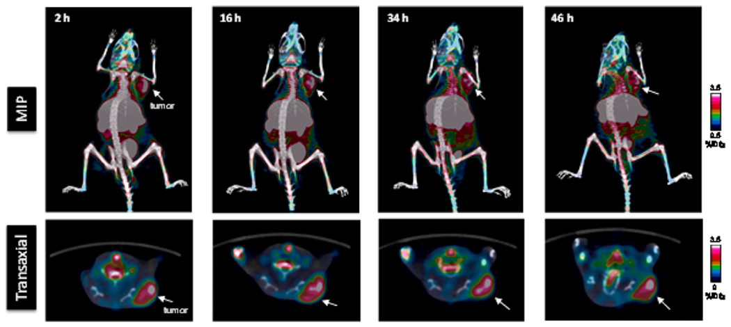
Representative PET/CT images of LNCaP tumor xenografts with 64Cu-BAVI in a mouse model at 2, 16, 34, and 46 h p.i. Images are shown by maximum intensity projection (MIP). The tumors are indicated by white arrow.

Representative PET/CT images of LNCaP tumor xenografts with 64Cu-BAVI in a mouse model at 2, 16, 34, and 46 h p.i. Images are shown by maximum intensity projection (MIP). The tumors are indicated by white arrow.