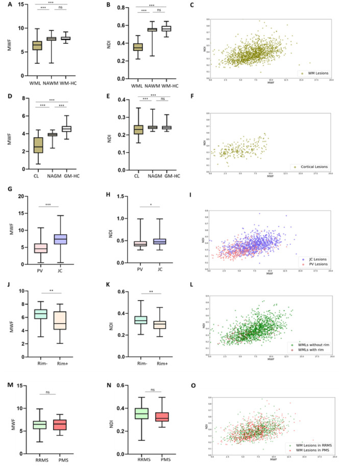
Figure 2.
MWF and NODDI-NDI in different types of multiple sclerosis lesions. (A and B) Box plots showing that MWF and NDI in white matter lesions (WMLs) is lower than MWF and NDI in NAWM and white matter in healthy controls (WM-HC). (C) NDI-MWF scatter plots in white matter lesions. (D and E) Box plots showing that MWF and NDI in cortical lesions (CL) is lower than in NAGM and grey matter in healthy controls (GM-HC). (F) NDI-MWF scatter plots in cortical lesions. (G and H) MWF and NDI are lower in periventricular (PV) lesions than in juxtacortical (JC) lesions. (I) NDI-MWF scatter plot in periventricular and juxtacortical lesions. (J and K) MWF and NDI are lower in Rim+ lesions than in Rim− lesions. (L) NDI-MWF scatter plot for white matter lesions with and without paramagnetic phase rim on both 3D EPI unwrapped phase and 3D EPI QSM images. (M and N) MWF and NDI were not different between RRMS and PMS. (O) NDI-MWF scatter plots for white matter lesions in RRMS versus PMS. NDI; NODDI-NDI. ***P < 0.0001; **P < 0.001; *P < 0.05. ns = not significant; WM = white matter.

