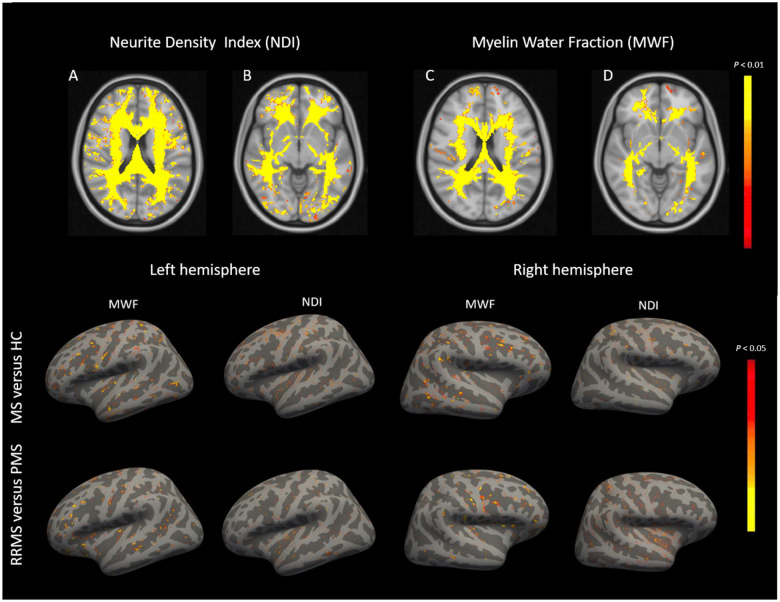
Figure 4.
Comparison of NDI and MWF in normal-appearing brain tissue between patients and controls. Top row: (A–D) Compared to healthy subjects (HC), multiple sclerosis (MS) patients show a widespread NODDI-NDI reduction in normal-appearing white matter and—to a smaller extent—a diffuse MWF reduction. Bottom row: There are patchy reductions in MWF and NODDI-NDI in the normal-appearing cortical surface of multiple sclerosis patients versus healthy controls and in PMS versus RRMS patients.

