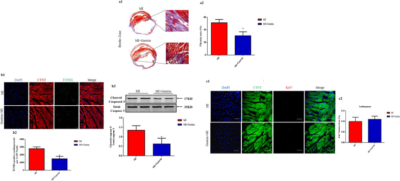
Fig. 3.
Gastrin decreases cardiomyocyte apoptosis and fibrosis after myocardial infarction. a Representative images (a1) and quantification (a2) of the fibrotic area in infarct border zone by Masson trichrome staining at 28 days post-MI. b Representative images (b1) and quantification (b2) of terminal deoxynucleotidyl transferase dUTP nick-end labeling–positive (TUNEL+) cardiomyocytes in infarct border zone at 2 days after MI. TUNEL+ nuclei are stained green, cardiac troponin T antibody (cTnT) is red, and 4'-6-diamidino-2-phenylindole (DAPI) is blue. b3 Western blot was used to measure the protein levels of Caspase 3 in the infarct border zone at 2 days after MI. Results are expressed as the ratio of cleaved caspased 3 to total caspase 3. c, Representative images (c1) and quantification (c2) of cardiomyocyte proliferation 28 days after myocardial infarction, through Ki67 immunostaining. Cardiac troponin T antibody (cTnT) is green, Ki67 is red, and 4'-6-diamidino-2-phenylindole (DAPI) is blue. a and c Scale bar = 50 µm. b Scale bar = 20 µm. n = 8. *P < 0.05, vs. MI mice. P = NS means no significant difference

