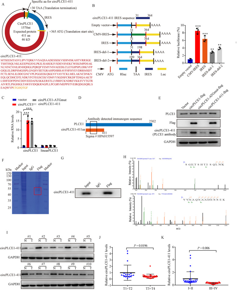
Fig. 3.
circPLCE1 encodes a 411 amino acid novel protein, circPLCE1-411. A Upper panel, the putative open reading frame (ORF) in circPLCE1. Lower panel, the sequences of the putative ORF encoded amino acid sequences are shown. B The putative IRES activity in circPLCE1 was tested. Left panel, IRES sequences in circPLCE1 or its different truncations were cloned between Rluc and Luc reporter genes with independent start and stop codons. Right panel, the relative luciferase activity of Luc/Rluc in the above vectors was tested, n = 3. C qRT-PCR analysis of circPLCE1 and PLCE1 linear mRNA expression in HCT8 cells transfected with empty vector, circPLCE1 vector, circPLCE1-ATGmut vector and circPLCE1-411 vector, n = 3. D Illustration of antibody detected immunogen sequence which could recognize both PLCE1 and circPLCE1-411 proteins. E Western blot analysis of PLCE1 and circPLCE1-411 protein levels in HCT8 cells transfected with empty vector, circPLCE1 vector, circPLCE1-ATGmut vector and circPLCE1-411 vector with indicated antibodies. F The lysates from immunoprecipitation assays were separated by SDS-PAGE. Protein bands near 50 kDa were excised manually and summited for identification by LC–MS/MS. G Western blot validation of circPLCE1-411 with anti-Flag and anti-PLCE1 antibody in immunoprecipitation products. H The identified circPLCE1-411 amino acids. I Western blot analysis circPLCE1-411 expression in paired CRC samples and normal adjacent tissues with indicated antibodies. J Comparison of circPLCE1-411 expression between patients with T stage 3–4 (n = 29) and those with T stage 1–2 (n = 21), detected by western blot. K Comparison of circPLCE1-411 expression between patients with clinical stage III–IV (n = 21) and those with clinical stage I–II (n = 29), detected by western blot. Values are represented as mean ± SD. **p < 0.01, ***p < 0.001, by 2-tailed Student’s t test (J and K) and one-way ANOVA (B and C)

