Fig. 4.
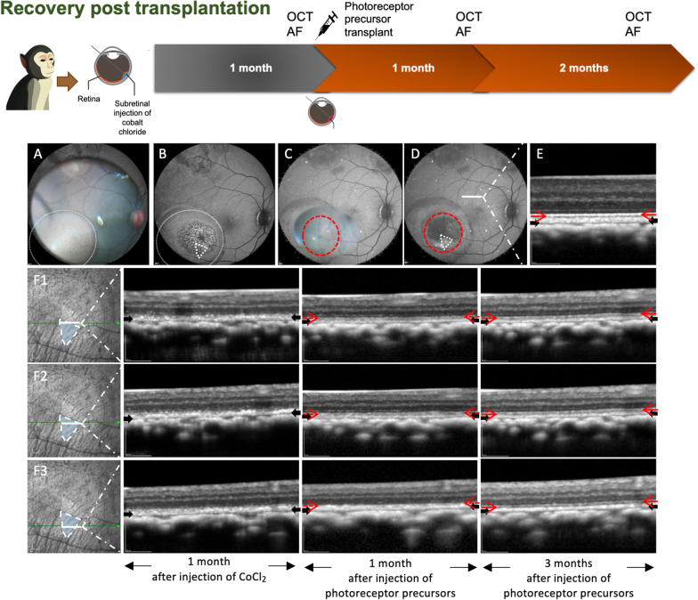
Retinal structure recovery in CoCl2 damaged zone by photoreceptor precursors injection. A Intraoperative imaging of CoCl2 injection superimposed on fundus autofluorescence (FAF) image. The site of CoCl2 was within the white dotted circle. B FAF imaging 1 month post-CoCl2 injection and immediately prior to photoreceptor precursors transplantation. C Intraoperative imaging of retinal photoreceptor precursors injection superimposed on the FAF image. The site of photoreceptor precursors was demarcated within the red dotted line. D FAF imaging was performed 4 months post-CoCl2 injection and 3 months post-photoreceptor precursors injection. The area outlined by the white dotted triangle was selected for analysis of morphological changes. The autofluorescence signal intensities in this zone reverted to normal suggesting recovery of RPE function. E OCT scan showed normal retinal structure outside the CoCl2 injected zone. The red and black arrows indicate the intact ellipsoid zone and RPE/Bruch’s membrane complex, respectively. F1 to F3 tracked OCT line scans of three different regions within the photoreceptor precursors injected zone. The ellipsoid zone was indistinct at 1 month post-CoCl2 injection but demonstrated progressive recovery with near-complete restoration at 3 months post-photoreceptor precursors injection

