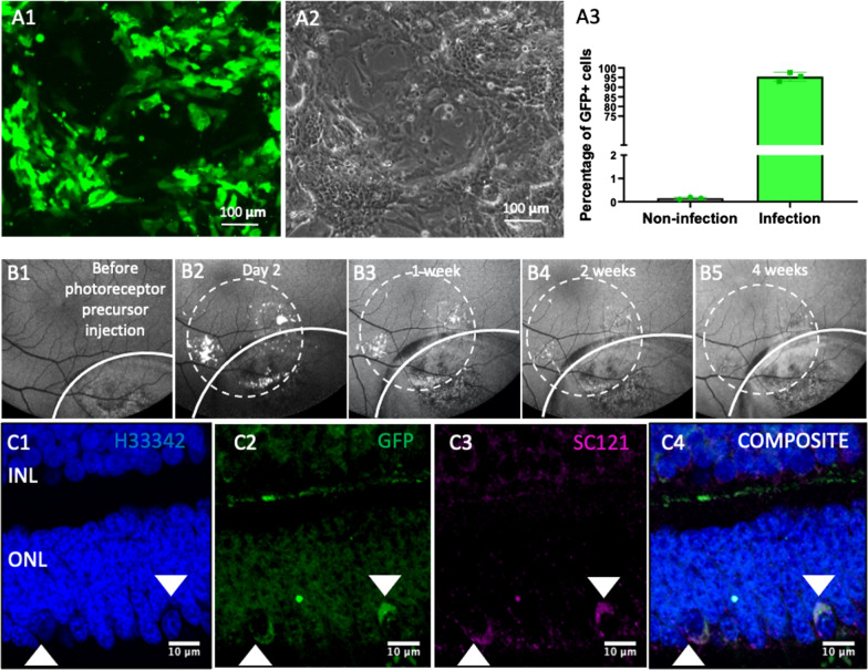
Fig. 5.
GFP-labelled photoreceptor precursors culture and in vivo tracking post-transplantation in NHP. A1 Representative fluorescence micrograph for GFP labelling by pCDH-GFP lentivirus injection. A2 Phase contrast image of the cells in A1. A3 Quantification of GFP labelling efficiency (n = 3) by flow cytometry. 95% of the cells were labelled. B1–B5 fundus autofluorescence images of in vivo follow up of transplanted photoreceptor precursors. The white curved lines indicated the region injected with CoCl2. The white dotted circles indicated the region injected with GFP-labelled photoreceptor precursors suspension. C1 to C4 Immunofluorescence staining of paraffin embedded tissues collected 12 weeks post-photoreceptor precursors transplant show GFP-positive (C2, white arrows) and SC121-positive (C3, white arrows) cells in the outer nuclear layer (ONL). The GFP- and SC121-positive cells co-localize (C4, white arrows), indicating that they are transplanted human photoreceptor precursors. Scale bars, 100 µm in A1 and A2, 200 μm in B1 to B5, 10 μm in C1 to C4

