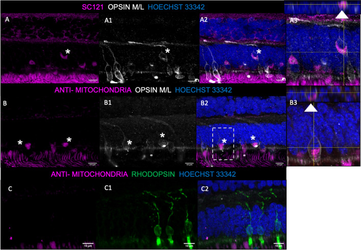
Fig. 6.
Survival and differentiation of transplanted photoreceptor precursors in the host NHP retina. A–A3, Anti-human SC121-positive cells appeared to survive in the outer nuclear layer (ONL) of the host NHP retina (A & A2, magenta). These cells co-localized with opsin M/L-positive staining (A2, grey, asterisk), suggestive that the transplanted photoreceptor precursors are able to differentiate into cones. Z-stack projection show the markers localized to a single cell (A3, white arrow). B–B3, Anti-human mitochondrial antibody (AMA)-positive photoreceptor precursors appeared to survive in the ONL of the host NHP retina (B & B2, magenta). They also co-localized with opsin M/L-positive cone photoreceptors (B2, grey, asterisk). Z-stack projection showed clear co-localization between AMA and opsin M/L around a nucleus (B3, white arrow). C–C2, AMA-positive photoreceptor precursors (C & C2, magenta) did not co-localize with rhodopsin-positive rod photoreceptors (C2, green). Nuclear staining with Hoechst 33342 (A2, B2, C2, blue) was used to identify the inner and outer nuclear layers. The cells not highlighted with an asterisk are most likely native NHP opsin M/L-positive cones. Scale bars: 10 μm

