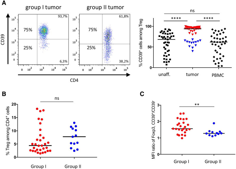
Fig. 2.
CD39 expression by Treg from colon adenocarcinoma patients. Frequencies of CD39+ Treg among total Treg, were determined by flow cytometry in single cell suspensions from tumors, unaffected colon tissue and peripheral blood. Colon tumors were divided into two groups based on CD39 expression by isolated intratumoral Treg, i.e. group I tumors (≥ 75% of CD39-expressing cells among intratumoral Treg) and group II tumors (< 75% of CD39-expressing cells among intratumoral Treg). A Representative FACS-plots, depicting CD39 expression by Treg from one group I tumor and one group II tumor, and a compilation of individual data points from the different locations (n = 40–44), with tumor-infiltrating Treg color-coded with red (group I) and blue (group II). B Lymphocytes isolated from group I and II tumors were analyzed for frequencies of total Treg among intratumoral CD4+ T cells. C MFI of Foxp3 expression (n = 36), shown as the MFI ratio of Foxp3 expression, between CD39+ and CD39− Treg in patients belonging to group I and II. Symbols represent individual values and horizontal lines the median. Individual values are color-coded in red (group I) and blue (group II). **p < 0.01, ****p < 0.0001

