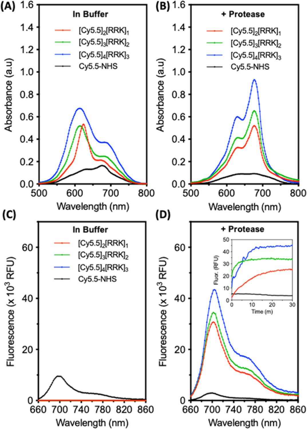
Figure 3.

Proteolysis of cyanine–peptide conjugates in the buffer induces a red shift and fluorescent activation. (A) Absorbance spectra of free dye, dimer, trimer, and tetramer conjugates at 7 μM in buffer (10 mM NH4HCO3, 1% DMSO, and pH 8.0). (B) Absorbance spectra of the molecules after incubation with 5 μM trypsin (30 min, 37 °C). (C) Fluorescence spectra of the molecules in the buffer (ex: 600 nm); inset: kinetic monitoring of fluorescence after the addition of 5 μM trypsin. (D) Fluorescence spectra after 30 min of incubation with trypsin. Inset: fluorescence activation from 0 to 30 min.
