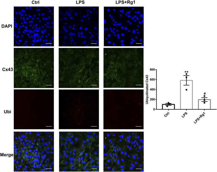
FIGURE 9.
Rg1 reduces ubiquitinated Cx43 in LPS-treated glial cells. The confocal microscopy images showed double-stained with antibodies against Cx43 (green) and ubiquitin (red). Nuclei were labeled with blue. Scale bar = 150 μm. n = 3. **p < 0.01, compared with the Ctrl group; # p < 0.05, compared with the LPS group.

