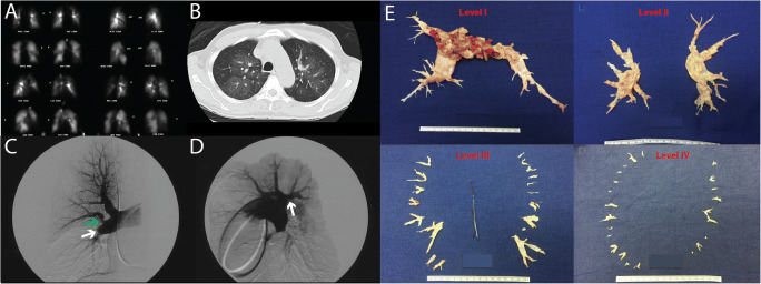
Figure 2.
Imaging and thrombus in CTEPH. A V/Q scan in the same patient showing multiple bilateral mismatched defects. B CT scan showing clear vascular mosaicism. C The white arrow highlights an angiographic “pouch” occlusion of the right interlobar vessel. The presence of organized thromboembolic disease is also evident by “web” narrowing of the proximal anterior upper lobe artery (green arrow). D The lateral right pulmonary arteriogram in the same patient shows another “web” narrowing (white arrow) of the proximal posterior upper lobe vessel not appreciated on the AP films. (The figure was published in Fedullo PF, Auger WR. “Medical Management of the Thoracic Surgery Patient”, 2010 pp: 477-482, copyright Elsevier [2010]) [37]. E University of California San Diego classification of PEA disease levels with illustrative figures for each level (Reprinted with permission of the American Thoracic Society. Copyright © 2021 American Thoracic Society. All rights reserved. Madani M, M. E. Ann Am Thorac Soc, 2016, 13 Suppl 3, S240-S247. Annuals of the American Thoracic Society is an official journal of the American Thoracic Society.) [42].

