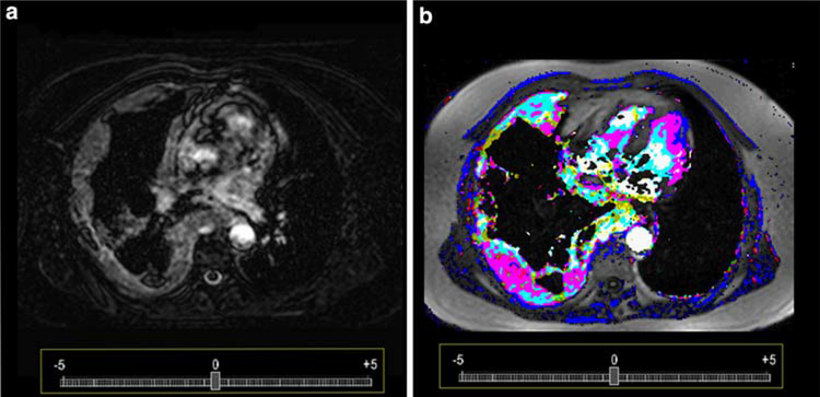
Fig. 1.
Subtraction images (a) presenting a patient with malignant pleural mesothelioma adjacent to the right chest wall. The high masculinity of the tumor can be already appreciated to most of the tumor tissue. Color code display (b) presents the same tumor vasculature as in a, but on the basis of a post processed dynamic contrast enhanced MRI. Tumor vasculature is better visualized and hyper- and hypo-vascular areas can be well differentiated

