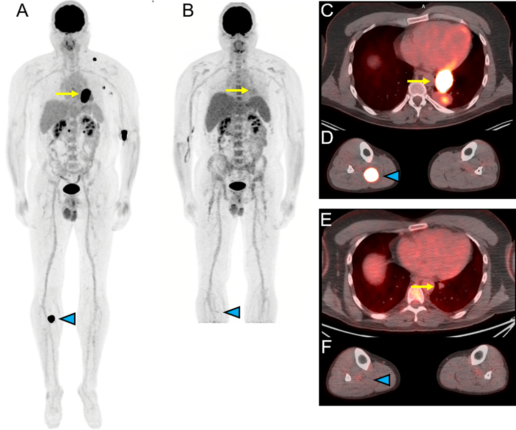
Figure 1.
18F-FDG-PET/CT for assessing response to immune checkpoint therapy (ipilimumab/nivolumab) in a 44 year-old-man with metastatic melanoma of unknown primary. Selected images prior to therapy are shown in panels A (maximal intensity projection (MIP) image), C and D (fusion images of the chest and lower extremities) while selected images obtained approximately 4 months later after starting therapy are shown in panels B (MIP), E and F (fusion images of the chest and lower extremities). The MIP images (A, B) demonstrate resolution of increased FDG uptake associated with multiple metastases with the fusion images demonstrating response in a left lower lobe mass (yellow arrow, C and E) and in a right proximal calf lesion (blue arrowhead, D and F). Courtesy of Jonathan McConathy, MD, PhD, at the University of Alabama at Birmingham (UAB).

