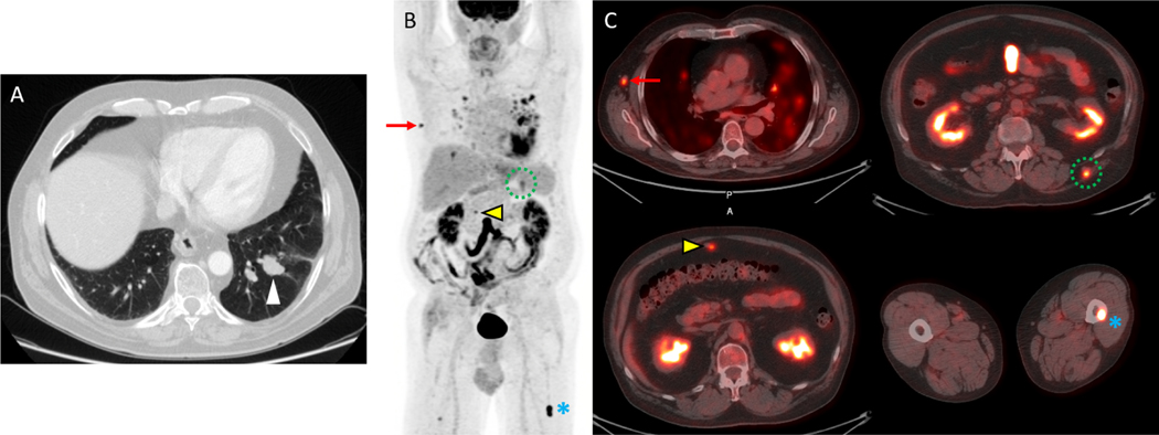
Figure 2.
18F-FDG-PET/CT detects multiple metastases in a 71 year-old-man with recurrent melanoma. A) An axial imaging from a diagnostic chest CT demonstrates a suspicious left lower lobe nodule which was subsequently biopsied bronchoscopically and shown to be a melanoma metastasis. The remainder of the diagnostic CT examination of the chest, abdomen and pelvis demonstrated an equivocal omental nodule but no definite metastatic disease. Based on these results, the patient began evaluation for a wedge resection of this metastasis which included restaging with FDG PET/CT. B) The maximal intensity projection (MIP) imaging from an FDG-PET study demonstrates multiple metastases as well as inflammation in the lungs. C) Fused PET/CT images demonstrate metastases with increased FDG uptake in the right axilla (red arrow), omentum (yellow arrow), left back musculature (dotted green circle) and left femur (blue asterisk). Surgical biopsy confirmed the right axillary lymph node metastasis, and the patient was treated with systemic therapy rather than metastectomy. Courtesy of Jonathan McConathy, MD, PhD, at the University of Alabama at Birmingham (UAB).

