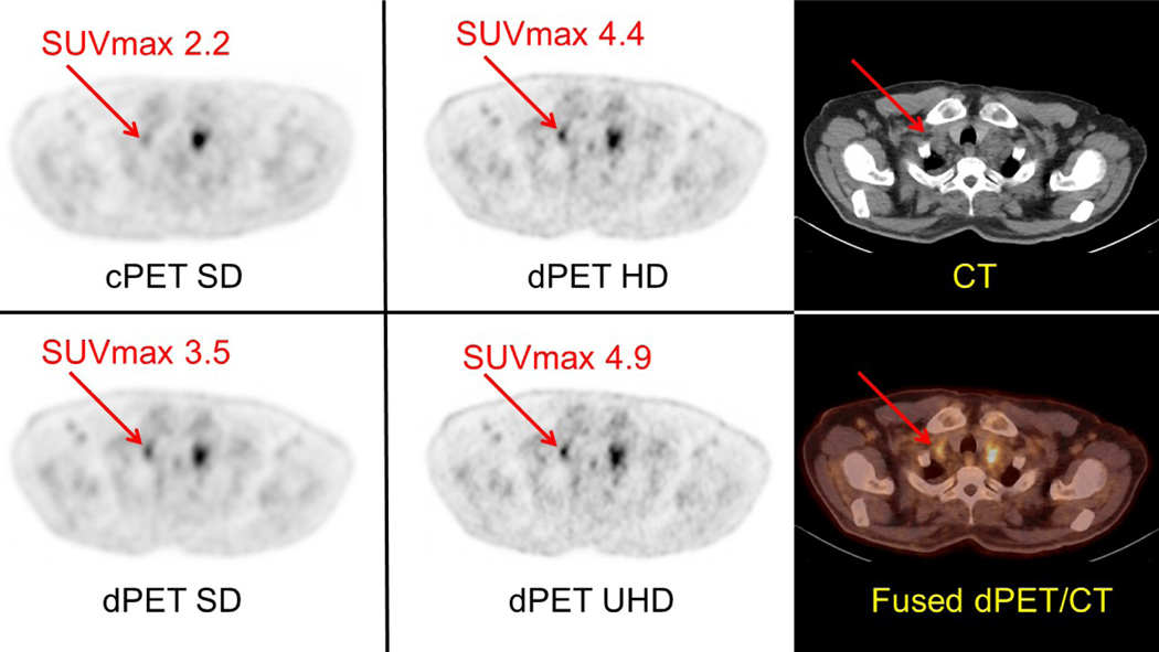
Figure 4:
Intra-individual comparison in a patient imaged using a cPET/CT (Gemini 64 ToF, Philips) system and a dPET/CT (Vereos, Philips) system with different reconstruction matrix/voxel volume sizes. This case further demonstrates the capabilities of higher definition dPET reconstructions to reduce partial volume effects, more precisely localize FDG activity especially within small lesions, and increase the visual conspicuity of FDG-avid lesions. In addition, higher definition dPET reconstructions enable more precise measurement of SUVmax in small lesions (i.e., <15 mm in short axis). The patient was intravenously administered a standard dose of 478 MBq of FDG and then underwent imaging on the dPET/CT system at 53 minutes and the cPET/CT system at 81 minutes post injection. Both cPET and dPET emission scans were acquired with 90 seconds per bed position. Left and Middle: Axial images taken at the level of a right supraclavicular lymph node (red arrows) are shown with associated SUVmax value. Although there is a FDG-avid soft tissue lesion mass noted in the left supraclavicular region on both cPET and dPET, there is a small lymph node in the right supraclavicular regions (red arrow) which is visually more conspicuous on dPET images when compared with cPET and becomes more suspicious with higher definition dPET reconstructions. Right: Corresponding attenuation correction CT image and fused dPET/CT image at the level of the right supraclavicular lymph node.

