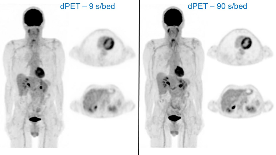
Figure 5:
Intra-individual comparison in a patient imaged using a dPET/CT (Vereos, Philips) system and acquired with different dPET image acquisition times (ie, standard = 90 seconds per bed position, and ultra-fast = 9 seconds per bed position). This case demonstrates the capabilities of dPET technology to facilitate ultra-fast PET imaging with markedly reduce PET image acquisition times (1/10th of the standard acquisition time) while generating visually comparable image quality. The patient was intravenously administered a standard dose of 484 MBq of FDG and then underwent ultra-fast imaging (LEFT - 9 seconds per bed with a total PET acquisition <2 minutes) on the dPET/CT system at 53 minutes post injection followed by standard imaging (RIGHT - 90 seconds per bed with a total PET acquisition ~16 minutes) at 57 minutes post injection. For each acquisition, a maximum intensity projection image from SD dPET using optimized reconstruction methodologies are shown along with representative axial dPET SD images from taken at the levels of the heart and liver. The ultra-fast whole body dPET image acquisition produced visually comparable image quality when compared with the standard whole-body dPET image acquisition. In addition, the physiologic FDG activity is qualitatively and quantitatively similar on the ultra-fast and standard dPET acquisitions at the levels of the heart and liver. FDG uptake in the normal liver has a SUV mean = 1.9 for both dPET acquisitions.

