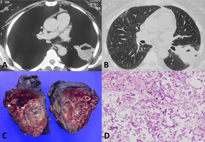
Fig. 1.
Computed tomography images of a patient with pulmonary mucormycosis (following COVID-19) showing a thick-walled cavity with an air-fluid level in the left lower lobe (A and B). Gross photograph (C) of the resected specimen showing cavity filled with necrotic material. Photomicrograph (D) demonstrating broad aseptate fungal hyphae conforming to the morphology of mucormycosis in a necrotic background (H&E, × 200)

