
Fig. 2.
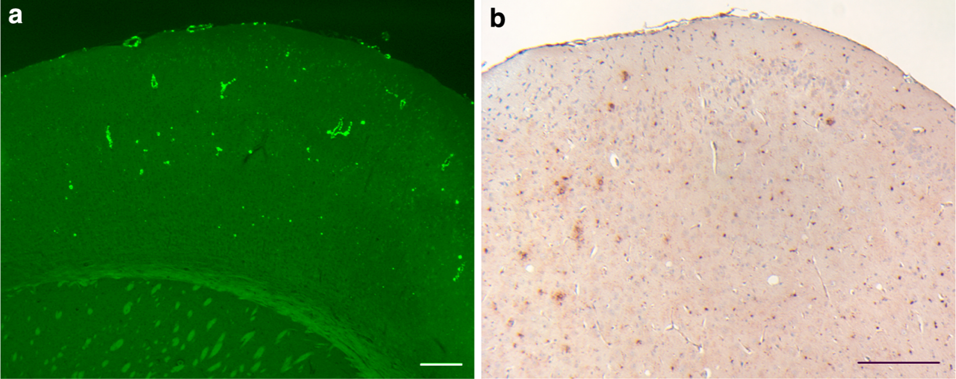
ADan deposition in the cerebral cortex. a All cerebral cortical layers of Tg-FDD mice show ThS-fluorescent amyloid deposition. b Amyloid deposits were immunoreactive with anti-ADan antibodies (Ab 1700), which also stained the subpial region of the cerebral cortex and diffuse parenchymal deposits. Sections were from a 21 month-old Tg-FDD mice. Staining with ThS (a). Immunohistochemistry using Ab 1700 was done as described (Vidal et al. 2009) (b). Scale bars a, b 200 μm