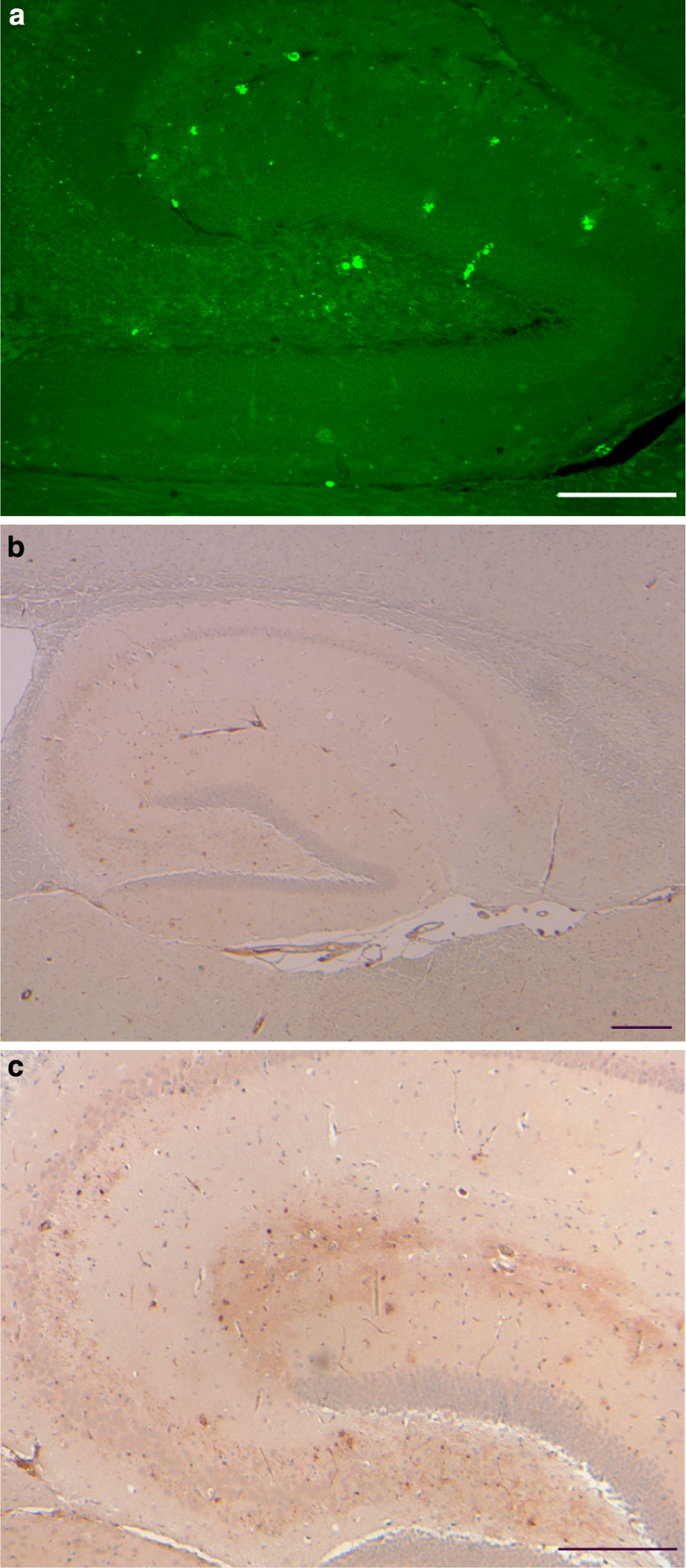
Fig. 3.

Vascular and parenchymal ADan deposition in the hippocampus. a Amyloid deposition was observed throughout the hippocampal formation of Tg-FDD mice by ThS. Fluorescent small, punctate deposits, ~3 μm in diameter were also observed. b ADan-immunopositive structures outlined the hippocampus. ADan was found in the walls of large and medium size vessels and in the walls of vessels of the hippocampal fissure. Sections were from a 21-month-old Tg-FDD mice. Staining with ThS (a). Immunohistochemistry using Ab 1700 (b, c). Scale bars a 100 μm, b, c 200 μm
