
Fig. 5.
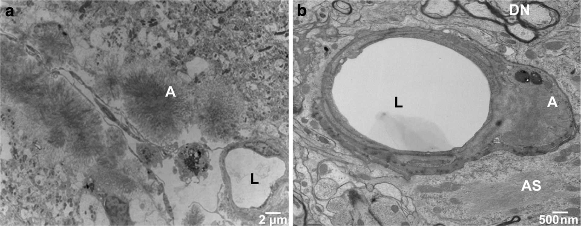
Ultrastructural analysis of cerebral vascular ADan deposition in Tg-FDD mice. a Amyloid deposits in leptomeningeal vessels of the cerebellum were observed within the vessel wall or appeared as circumscribed deposits with radially oriented fibrils. b Abnormal thickening of the basal lamina as well as degeneration and death of endothelial cells was observed in hippocampal vessel walls, in some cases with penetrance of amyloid into the vascular basement membrane. Transmission electron microscopy (TEM) of a 18-month-old Tg-FDD mouse. Amyloid (A), vessel lumen (L), dystrophic neurites (DN) and astrocytic processes (AS) were identified