
Fig. 7.
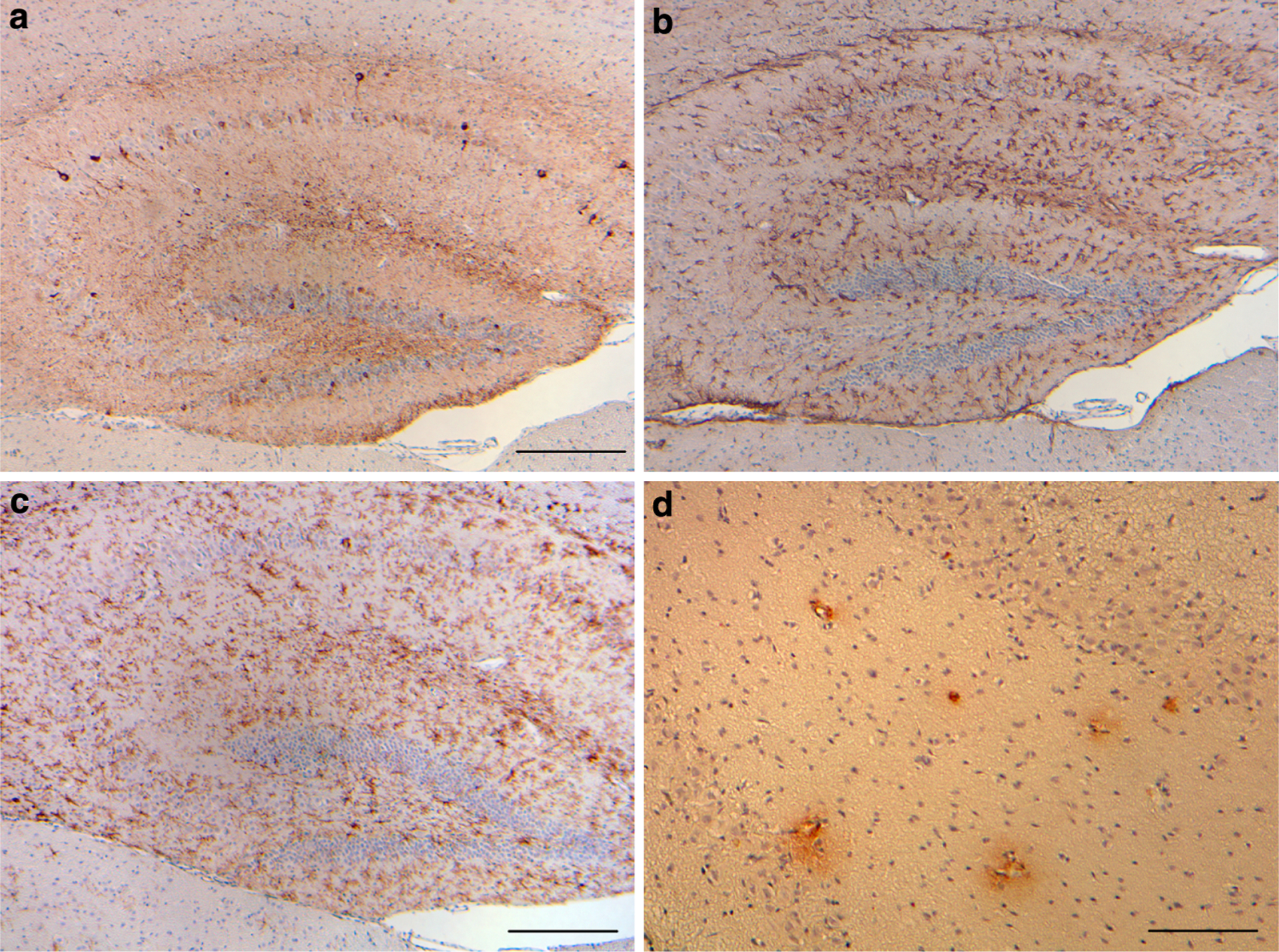
Tg-Tau × Tg-FDD mice. a Phosphorylated tau, b GFAP-positive reactive astrocytes, and c keratan sulfate-positive activated microglia in the hippocampus and motor cortex of Tg-Tau × Tg-FDD mice. d Antibodies against ADan immunolabel hippocampal blood vessels and perivascular deposits in Tg-Tau × Tg-FDD mice. Sections were from a 12-month-old homozygous Tg-Tau × Tg-FDD mice. Immunohistochemistry using AT8 (a), anti-GFAP (b), 5D4 (c), and anti-ADan (1700) (d). Scale bars a–c 200 μm, d 100 μm