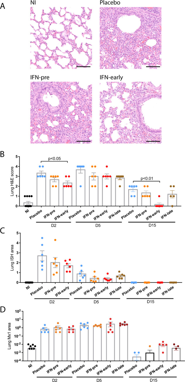
Fig 3. Histopathological analysis of the impact of type I IFN-α treatment.
(A) Representative pictures were selected to display the pathology from haematoxylin and eosin (H&E) stained lung section from animals at day 2 post infection. Scale bar: 100μm. (B) Severity of lung pathology based on lesional scores evaluated from haematoxylin and eosin (H&E) stained lung section. Statistical analysis: Mann-Whitney test. (C) Quantification of percent lung area positive for viral RNA in lung sections stained with RNAScope in situ hybridization (ISH). Statistical analysis: one-way ANOVA with Tukey’s multiple comparisons test. (D) Quantification of percent lung area positive for Mx1 protein detected by immunohistochemistry (IHC). Statistical analysis: one-way ANOVA with Tukey’s multiple comparisons test. D2: day 2 post infection; D5: day 5 post infection; D15: day 15 post infection. Results are expressed as means ± SEM.

