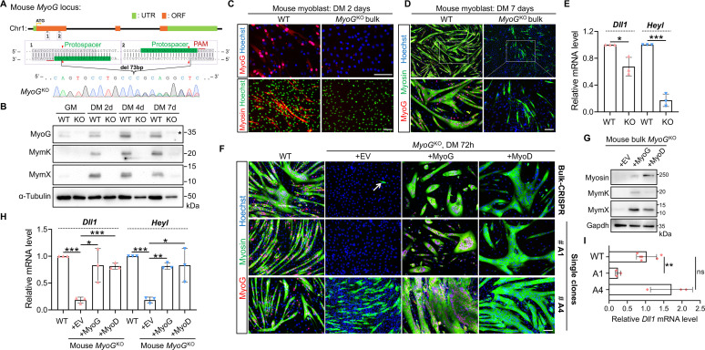
Fig 7. Responses of Dll1 expression upon deletion of MyoG from mouse myoblasts.
(A) Schematic of mouse MyoG gene structure and an example of Sanger sequencing result after CRISPR/Cas9 mediated gene editing in C2C12 mouse myoblasts. (B) Western blotting results to show the expression levels of myogenic markers in WT and MyoGKO myoblasts. Star highlights a non-specific band. (C, D) Immunostaining results of mouse WT and MyoGKO myoblasts at day 2 (C) and day 7 (D) post differentiation. (E) qPCR results of Dll1 and Heyl in WT and MyoGKO myoblasts. Cells were differentiated for 48 hours. n = 3. (F) Immunostaining result of MyoG and myosin for bulk CRISPR-treated myoblasts (top) and two isolated KO clones (bottom). Note that although MyoG were invariably depleted, the two KO clones showed a large variation of myogenic capacity. Cells were induced for differentiated for 72 hours. (G) Western blotting results to show the expression levels of myogenic differentiation markers in and bulk CRISPR-treated MyoGKO myoblasts. Cells were differentiated for 72 hours. (H, I) qPCR results for gene expression in WT and bulk CRISPR-treated MyoGKO myoblasts (H) or MyoGKO single-knockout clones (I). Cells were differentiated for 48 hours. n = 3. Data are means ± SEM. *P < 0.05, **P < 0.01, ***P < 0.001. ns, not significant. Scale bars, 100 μm.

