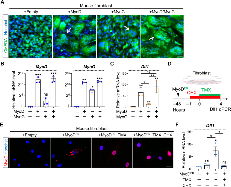
Fig 8. Sufficiency test of Dll1 inductions by MyoD in fibroblasts.
(A) Fluorescence images of cell cytosol dye CMFDA (green) to highlight fibroblast syncytia induced by MyoD or MyoG. Scale bar, 100 μm. (B, C) qPCR results of MyoD, MyoG (B), and Dll1 in mouse fibroblasts with forced expression of MyoD, MyoG or both. n = 3. (D) Schematic of experimental design to test the sufficiency of MyoD transcriptional activity in activating Dll1 expression in fibroblasts. (E) MyoD immunostaining of fibroblasts. TMX: tamoxifen. CHX: cycloheximide. Scale bar, 25 μm. (F) qPCR results of Dll1 expression in fibroblasts after treatment illustrated in D. Note that induction of Dll1 expression by MyoD was abolished upon CHX treatment (5 hours). n = 3. Data are means ± SEM. *P < 0.05, **P < 0.01, ***P < 0.001. ns, not significant.

