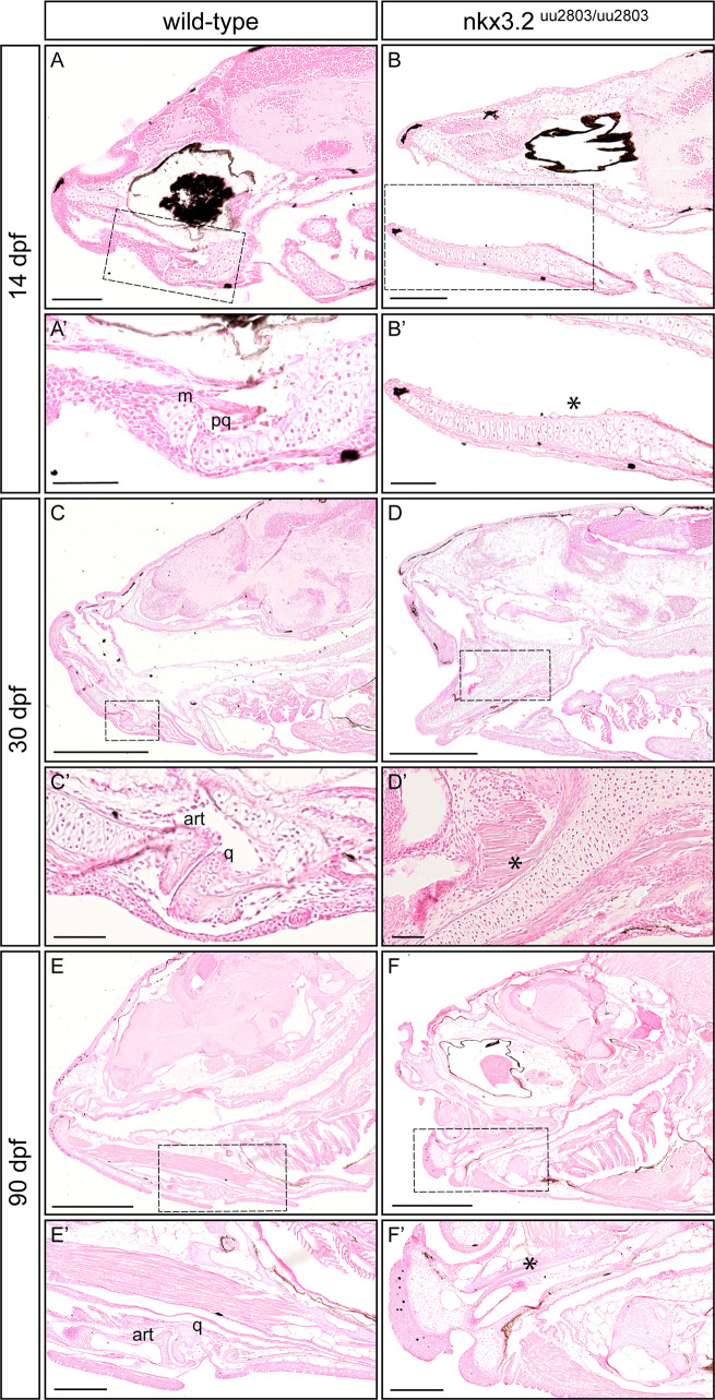
Fig 3. Histological analysis reveals loss of jaw joint without affected chondrocyte hypertrophy in the first pharyngeal arch of nkx3.2uu2803/uu2803 zebrafish.
Sagittal sections of wild-type 14 dpf (A, A’), 30 dpf (C, C’) and 90 dpf (E, E’) and nkx3.2 mutant 14 dpf (B, B’) 30 dpf (D, D’) and 90 dpf (F, F’) zebrafish stained with Nuclear Red. (A, A’, C, C’, E, E’) Stained histology sections displaying a normal jaw joint development between Meckel’s cartilage (m) and palatoquadrate (pq), respectively articular (art) and quadrate (q) in wild-type fish. (B, B’, D, D’, F, F’) nkx3.2 mutants do not display a jaw joint. Chondrocyte maturation and ossification seem not to be affected in nkx3.2uu2803/uu2803 besides the absence of joint-typical cells lining the articulating elements. Dashed box in (A-F) marks the magnified region in (A’-F’). Asterisk marks the fused element. m–Meckel’s cartilage, pq–palatoquadrate, art–articular, q–quadrate. Scale bars: 200μm (A-F), 100μm (A’-F’).

