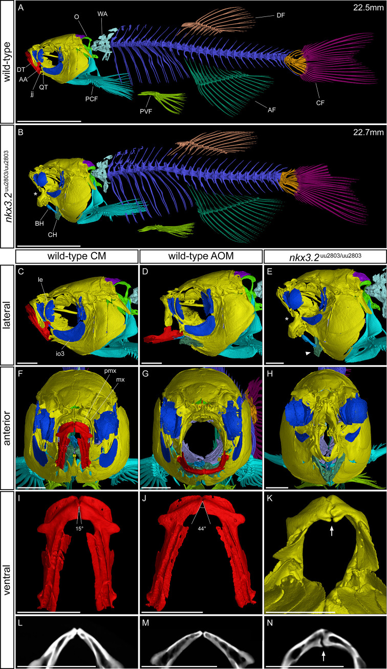
Fig 6. μCT reveals craniofacial phenotypes in adult wild-type and nkx3.2uu2803/uu2803 zebrafish.
(A, B) lateral view of wild-type and nkx3.2 mutant zebrafish at 90 dpf. (C, D, E) lateral view of the head of wild-type with closed mouth (CM), wild-type with artificially open mouth (AOM), and nkx3.2 mutant. (F, G, H) Anterior view of wild-type with CM, wild-type with AOM, and nkx3.2 mutant. (I, J, K) ventral view of isolated wild-type dentary (CM), wild-type dentary (AOM), and nkx3.2 mutant dentary. (L, M, N) Virtual thin sections (60μm thick, averaged) through the symphysis of the dentaries displayed in I, J, K, respectively. The measurements in mm refer to standard length (SL). Asterisks in (B, E) indicate the fixed open mouth phenotype caused by fused jaw joint. The arrowhead in (E) indicates the downturned ceratohyal phenotype relative to (D). Angles in (I, J) show the flexibility of the symphysis in wild-types, the arrow in K and N indicates the thickened and deformed symphysis phenotype in nkx3.2 mutants. Colour scheme: Red–lower jaw; dark blue–infraorbitals; dark green–ceratohyal and anal fin; blue–basihyal; lilac–branchial arches; dark purple–supraoccipital; lime green–exoccipital and basioccipital; yellow–all remaining craniofacial bones; cyan–cleithrum and pectoral fins, arctic blue–Weberian apparatus; blue violet–vertebrae; green–pelvic fins; bronze–dorsal fin; orange–caudal fin vertebrae and hypurals; magenta–caudal fin rays. AA–anguloarticular, AF–anal fin, BH–basihyal, CF–caudal fin, CH–ceratohyal, DF–dorsal fin, DT–dentary, io3 –infraorbital 3, jj–jaw joint, le–lateral ethmoid, mx–maxilla, O–occipital region, PCF–pectoral fin, pmx–premaxilla, PVF–pelvic fin, QT–quadrate, WA–Weberian apparatus. Scale bars: 5mm (A, B), 1mm (C-N).

