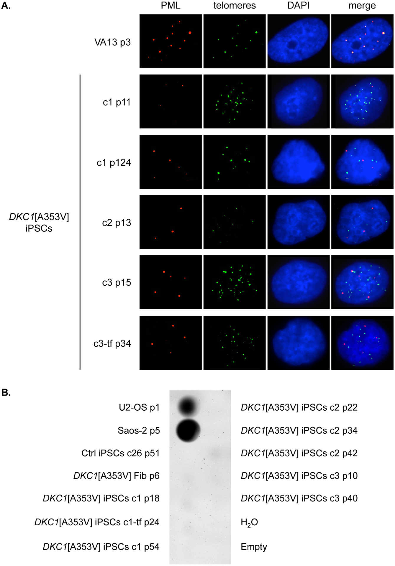
Figure 3 – Absence of alternative lengthening of telomeres in DKC1[A353V] iPSCs.

(A) Immunofluorescence of DKC1[A353V] fibroblasts (Fib p5) and iPSCs clones 1, 2, 3, and 3-tf at indicated passages (p). PML bodies were stained with an anti-PML antibody (red), and telomeres were labeled with FITC-conjugated PNA probe (green). Interphase nuclei were counterstained with DAPI (blue). Colocalization of PML bodies and telomeres was detected only in VA13 cells (positive control). (B) Dot blot of C-circle assay performed on genomic DNA from DKC1[A353V] fibroblasts (Fib p6) and iPSCs clones 1, 1-tf, 2, and 3 at indicated passages (p), as well as iPSCs from control (Ctrl iPSCs clone 26). Genomic DNA from the ALT-positive cell lines U2-OS and Saos-2 was set as positive control.
