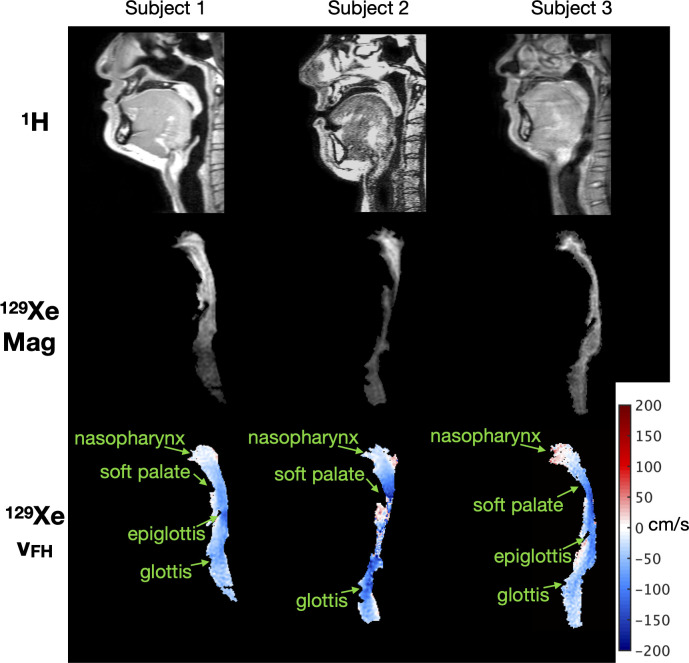
Fig 2. Example of MRI data acquired from each of the three subjects.
Top row: Sagittal 1H MRI of the head and neck anatomy including the upper airway, captured to create the virtual airway surface for CFD simulations. Middle row: Sagittal magnitude (“Mag”) MR images of inhaled 129Xe in the upper airway at a selected dynamic time (note: slices do not perfectly correspond to 1H MRI slices above). Bottom row: Foot-Head velocity (vFH) maps corresponding to the magnitude images above (one dynamic image), reconstructed from 129Xe gas PC MRI data. Red velocities represent flow in the foot to head direction, blue represents flow in the head to foot direction.

