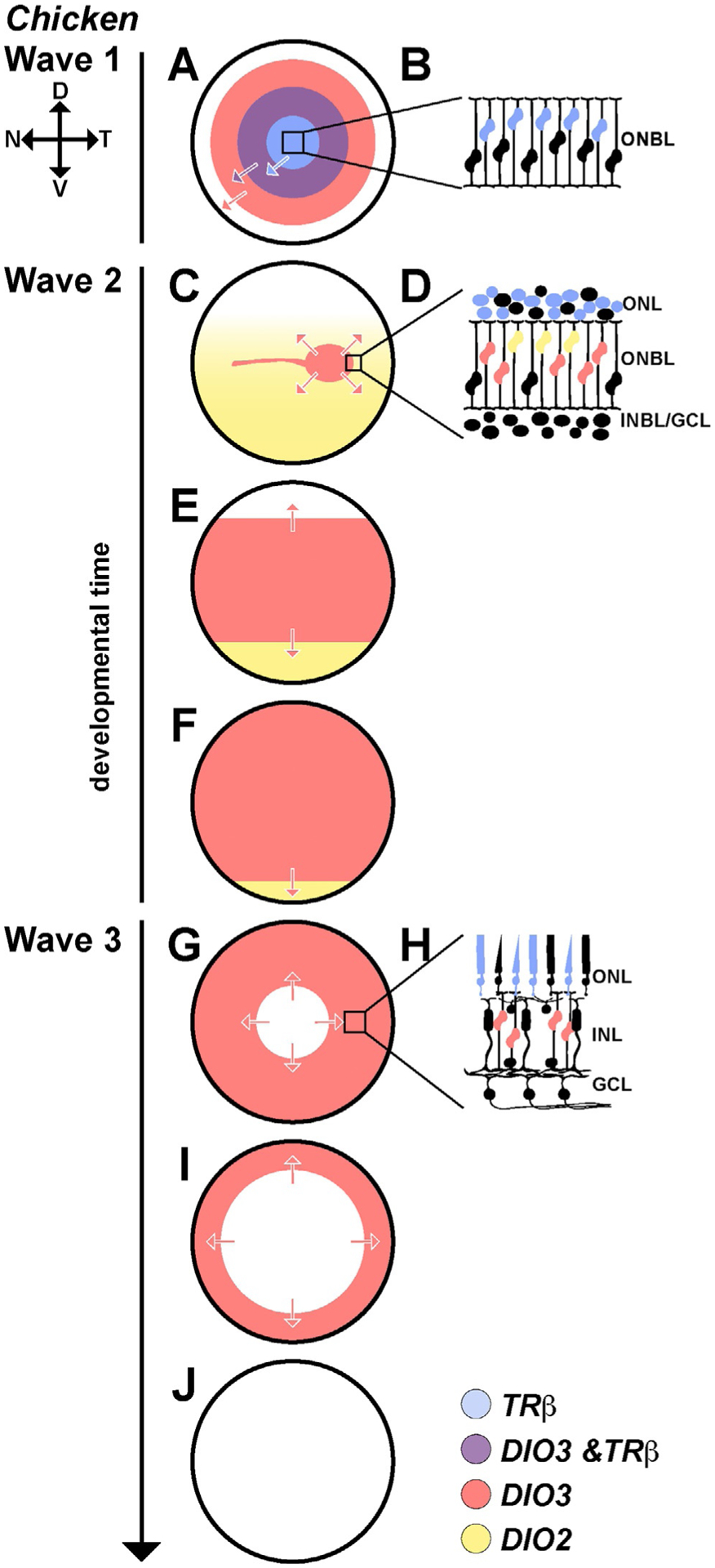
Fig. 4.

Thyroid hormone regulators are expressed in waves in the developing chicken retina. (A–J) All three waves move from center to periphery during development. (A) In wave 1, the front of the wave is led by DIO3 (red). Slightly delayed/behind DIO3 expression is TRβ (blue). At the transition, there is a region of overlap (purple) between the DIO3 and TRβ waves. (B) TRβ is expressed in a subset of progenitors, mostly in those closest to the developing photoreceptor layer. DIO3 is expressed in all cells in the wave. (C–F) In wave 2, DIO3 expression is limited to a strip running across the central retina (red), and DIO2 is expressed in the ventral retina and lowly expressed in the dorsal retina (yellow). During wave 2, this strip of DIO3 expands out toward the periphery as DIO2 expression becomes limited to the ventral periphery. (D) TRβ is expressed in developing photoreceptors (blue). DIO2 is expressed in a subset of cells in the upper ONBL (yellow). DIO3 is expressed in RPCs (red). (G, I and J) In wave 3, DIO3 expression (red) is lost from the center to the periphery, corresponding with the birth of Müller glia. (H) TRβ (blue) remains active in a subset of photoreceptors. DIO3 expression (red) is limited to the remaining RPC population. Adapted from Trimarchi, J. M., Harpavat, S., Billings, N. A., & Cepko, C. L. (2008). Thyroid hormone components are expressed in three sequential waves during development of the chick retina. BMC Developmental Biology, 8, 101. https://doi.org/10.1186/1471-213X-8-101.
