. 2021 Aug 19;10:e69160. doi: 10.7554/eLife.69160

Figure 5. GIV is required for fertility in male mice.

(A) Schematic showing the workflow for fertility studies in conditional GIV-cKO mice. After intraperitoneal injection of tamoxifen, male mice were first primed in two phases—first by co-housing with female littermates × 3 weeks, and subsequently by co-housing with female mice from Jackson laboratory (JAX) while the females acclimatized to the animal facility. The final ‘test’ group consisted of tamoxifen-injected WT and GIV-cKO male mice randomly assigned to and co-housed with three female mice from JAX, each with proven ability to get pregnant. (B–D) Confirmation of GIV-cKO in the mice after tamoxifen injection by genotyping (B), qPCR of testis tissues (C), and immunoblotting of testis lysates (D) (Figure 5—source data 1). (E) Kaplan–Meier plot showing the cumulative probability of pregnancy (expressed as %) in the females co-housed with either WT or GIV-cKO males. Statistical significance was assessed using log-rank analysis. *p<0.05 (Figure 5—source data 2). (F, G) Bar graphs showing the average litter size (F; Figure 5—source data 3) and sperm count (G; Figure 5—source data 4) in WT and GIV-cKO males. See also Figure 5—figure supplement 1 for quantifications of tested weight and length. (H) Immunohistochemistry staining on mouse testis. Scale bar = 200 µm. (I) Unsupervised clustering of WT and KO testis samples based on gene expression. Differentially expressed genes (DEGs) that were up- or downregulated in KO are annotated on the right side (Figure 5—source data 5). (J, K) Reactome pathway analyses showing the pathways that are up or downregulated in KO testis. (L) Summary of the most prominent conclusions from RNA-seq dataset.

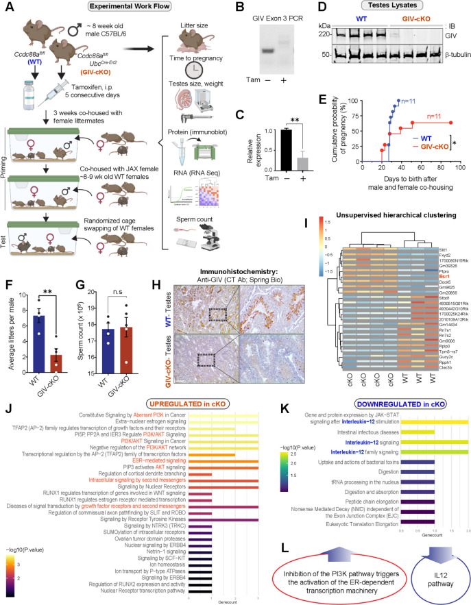
Figure 5—source data 1. Full-length, uncropped immunoblots on testes lysates with GIV and tubulin antibodies (corresponds to Figure 5D).
Figure 5—source data 2. Excel sheet with time to live birth values observed in females co-housed with WT and GIV-cKO mice (corresponds to graphs in Figure 5E).
Figure 5—source data 3. Excel sheet with litter size values from WT and GIV-cKO mice (corresponds to graphs in Figure 5F).
Figure 5—source data 4. Excel sheet with sperm count values from WT and GIV-cKO mice (corresponds to graphs in Figure 5G).
Figure 5—source data 5. Excel sheet with differential expression analysis-derived reactome pathway analyses of the most significantly up- and downregulated genes in GIV-cKO mice (corresponds to graphs in Figure 5I).

Figure 5.

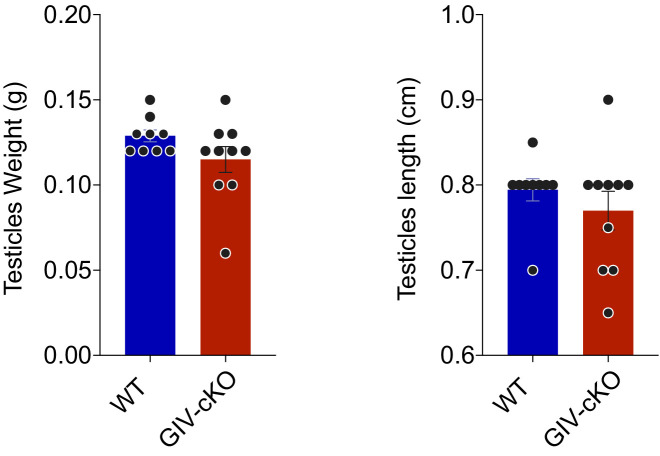
Figure 5—figure supplement 1. GIV is required for male fertility, but its depletion does not impact testes weight or length.

Figure 5—figure supplement 1.

GIV was depleted in male mice by intraperitoneal injection of tamoxifen (see Figure 5A). Bar graphs show the testes weight (left; Figure 5—figure supplement 1—source data 1) and length (right; Figure 5—figure supplement 1—source data 2) in WT and GIV-cKO males. All results are presented as average ± SEM. Statistical significance was assessed using unpaired t-test, non-significant p-values>0.05.
Figure 5—figure supplement 1—source data 1. Excel sheet with testes weight values from WT and GIV-cKO mice (corresponds to the left-side graph in Figure 5—figure supplement 1).
Figure 5—figure supplement 1—source data 2. Excel sheet with testes length values from WT and GIV-cKO mice (corresponds to the left-side graph in Figure 5—figure supplement 1).