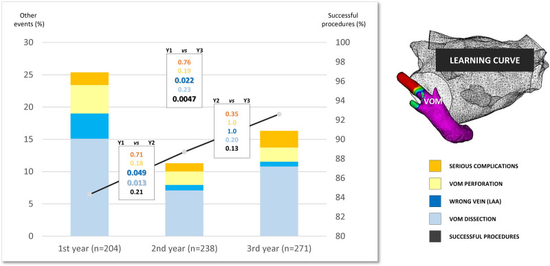
Figure 4.
Learning curve. Evolution of key procedural events over the years: successful vein of Marshall (VOM) ethanol infusion (black line), VOM dissection (pale blue), wrong vein ethanol infusion (dark blue), VOM perforation (yellow), and serious complications (orange). Statistical comparison with a P value is shown between each year (black-framed rectangle) and for each item (following identical color code). LAA indicates left atrial appendage.

