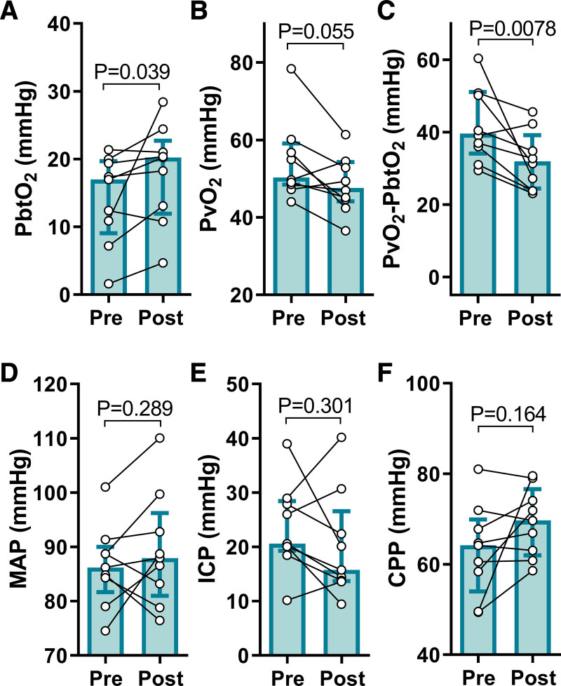
Figure 6.
Hypertonic saline increases brain tissue oxygen tension in post–cardiac arrest patients. Patients with hypoxic ischemic brain injury (HIBI) exhibiting brain hypoxia (HX, n=9) are denoted by the cyan coloured bars (median±interquartile range). Individual patient data are overlaid. The 1-h mean immediately pre hypertonic saline infusion (Pre) and the 6-h mean immediately following infusion (Post) are depicted for brain tissue Po2 (PbtO2; A), partial pressure of jugular bulb venous oxygen (PvO2; B), the PvO2-PbtO2 gradient (C), mean arterial pressure (MAP; D), intracranial pressure (ICP; E), and cerebral perfusion pressure (CPP; F). Hypertonic saline infusion increased PbtO2 (P=0.039) and decreased the PvO2-PbtO2 gradient (P=0.0078) in the patients with hypoxic HIBI. These changes occurred despite no alteration in the systemic hemodynamic input to the brain as indicated by unaltered MAP (P=0.289), ICP (P=0.301), and CPP (P=0.164). Only 4 patients with normoxic HIBI received hypertonic saline based on clinical indication, thus while the data are presented for transparency in the supplement (Figure V in the Data Supplement), no statistical analyses were completed on these data. The hypoxic HIBI patient data were compared preto-post hypertonic saline with a Wilcoxon Signed Ranks test, with significance assumed at P<0.05.

