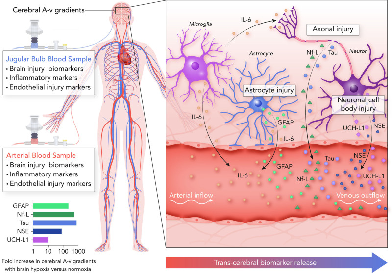
Figure 7.
Assessing secondary brain injury in post–cardiac arrest patients with hypoxic-ischemic brain injury through transcerebral biomarker release. This schematic outlines the measurement of serum biomarkers of brain injury in arterial and jugular venous blood samples to quantify transcerebral biomarker release (ie, the cerebral arterial-to-venous [A-v] gradient). This allows for experimental isolation of the cerebrovascular bed in the setting of an otherwise multisystem injury. The present investigation observed differences in the cerebral A-v gradients of markers of neuronal cell body (NSE [neuron-specific enolase] and UCH-L1 [ubiquitin carboxyl-terminal hydrolase L1]), axonal (Nf-L & Tau), and astroglial (GFAP [glial fibrillary acidic protein]) injury as well as neuroinflammation (IL-6 [interleukin-6]) between patients with secondary brain hypoxia compared with normoxia following a cardiac arrest. The difference in the magnitude of cerebral A-v gradients between patients with secondary brain hypoxia vs normoxia is demonstrated in the graph on the bottom left, with an accompanying illustration highlighting the sites of injury within the neurovascular unit on the right side of the figure.

