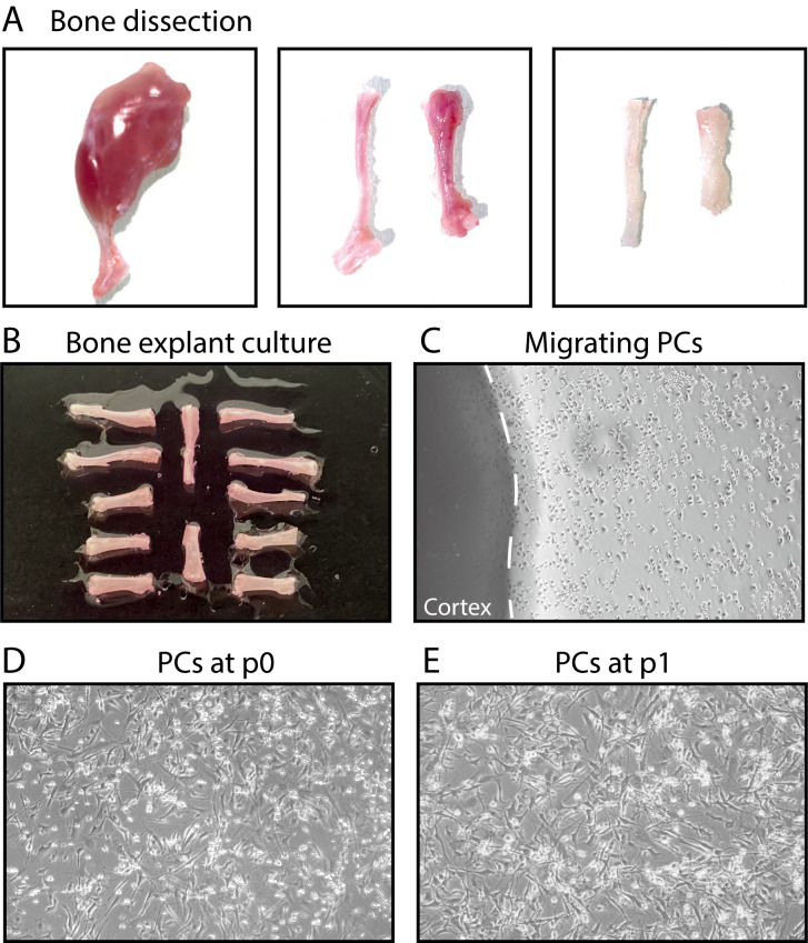
Figure 1. Steps of bone explant culture.
(A) Steps of bone dissection. Left, hindlimb free of skin. Middle, tibia, and femur free of muscle. Right, tibia and femur after cutting the epiphyses and flushing the bone marrow. (B) Tibias and femurs plated in a culture dish. (C) After a few days, periosteal cells (PCs) migrate out of the bone explants into the dish. (D-E) PCs at P0 and P1.

