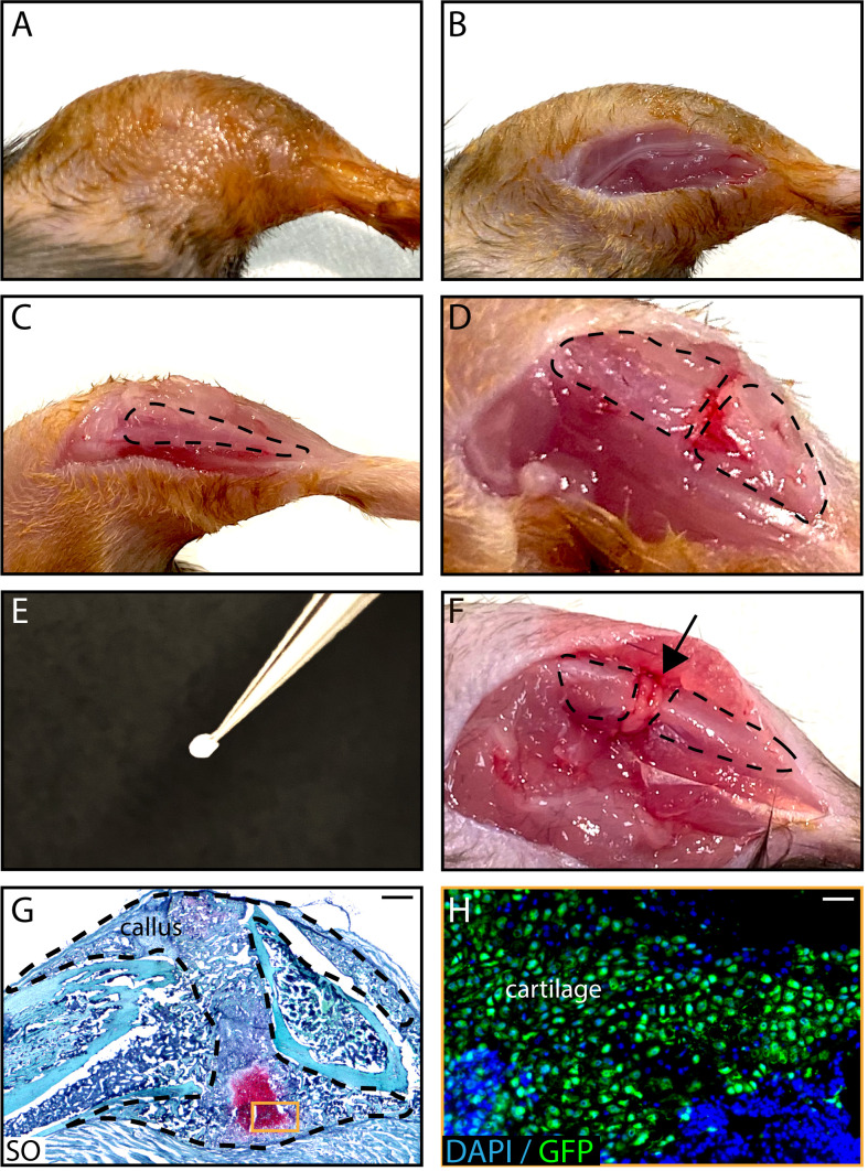
Figure 4. In vivo transplantation of PCs isolated from GFP-actin donors at the fracture site of wild-type hosts .
. After shaving and sanitizing the limb (A), an incision in the skin is performed (B) and the tibia is exposed (C, tibia is delineated by a dotted line) to induce a fracture (D). A Tisseel matrix pellet containing PCs (E) is transplanted at the fracture site (F, black arrow). (G) Representative image of a longitudinal callus section on day 14 post-fracture stained with SO. (H) GFP+ chondrocytes derived from PCs on an adjacent section of the callus. Scale bars: 1 mm (G) and 100 μm (H).

