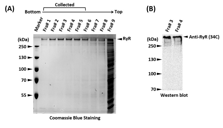
Figure 2. SDS-PAGE and western blot showing the sucrose gradient fractions and positive identification of the RyR.
A. SDS-PAGE gel stained with Coomassie Brilliant Blue of the sucrose gradient fractions (Fra#9 represents a mixture of all the remaining fractions). Fractions 1-6, containing pure RyR, were pooled. The unusual high molecular weight of RyR (565 kDa per monomer) places its band well above the top molecular weight marker, a distinguishing trait that in most scenarios suffices for positive identification. B. Western blot of fractions #3 and #4 of a sucrose density gradient using an anti-RyR antibody (34C) to confirm the identity of RyR.

