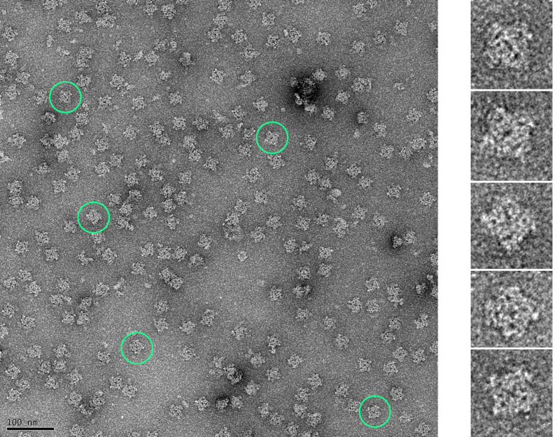
Figure 5. Micrograph of negatively stained RyRs purified from HEK293 cells (representative RyRs encircled in green).
Negative staining was performed to check the quality of the purified proteins using established protocols ( Ohi et al., 2004 ). Copper grids coated with a thin layer of carbon (CF300-Cu 300 mesh, Electron Microscopy Sciences) were first glow-discharged at 25 mA for 20 s. Purified RyRs (3.5 µl) were adsorbed onto the grid for 40 s, washed with deionized water, and stained with 0.75% uranyl formate; the excess stain was blotted off. Images were collected on an FEI Tecnai F20 TEM equipped with a GATAN Ultra Scan 4000 UHS (4k × 4k) camera at a magnification of 50,000×. Finer structural details of the RyR can be discerned in the enlarged images of individual RyRs corresponding to those highlighted in the micrograph.

